ES Journal of Case Reports
Multiple Carpometacarpal Joint Dislocation; a clinical image
Clinical Image
- Seyyed-Mohammad Qoreishi1, Hossein Mohebi2 and Seyyed-Mohsen Hosseininejad2, 3
- 1Assistant Professor of Orthopedic Surgery, Shahid Beheshti University of Medical Sciences, Iran
- 2Bone, Joint and Related Tissue Research Center, Shahid Beheshti University of Medical Sciences, Iran
- 3Golestan Rheumatology Research Center, Golestan University of Medical Sciences, Iran
- *Corresponding author: Seyyed-Mohsen Hosseininejad, Bone, Joint and Related Tissue Research Center, Akhtar Hospital, Shahid Beheshti University of Medical Sciences, Tehran, Iran
- Received: Feb 10, 2020; Accepted: Feb 14, 2020; Published: Feb 25, 2020
- Copyright: 2020 © Seyyed-Mohsen Hosseininejad. This is an open-access article distributed under the terms of the Creative Commons Attribution License, which permits unrestricted use, distribution, and reproduction in any medium, provided the original author and source are credited.
Traumatic fracture dislocations of second to fifth carpometacarpal (CMC) joints is a rare injury making up less than 1% of hand injuries [1]. Notifying the joint disruption could be difficult due to swelling and overlapping of bones. The mechanism of injury is usually due to high energy trauma e.g. car accidents. Dorsal CMC joint dislocations are more common than volar CMC joint dislocations. In literature, most cases presented single CMC joint dislocation, but few cases are available about multiple CMC joint fracture dislocation [2,3].
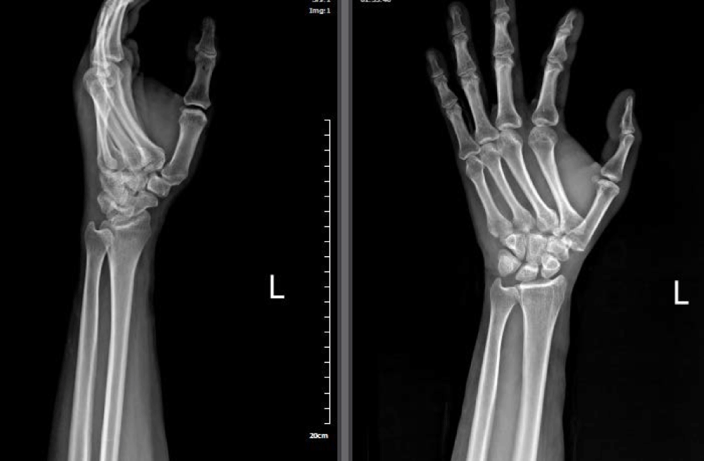
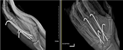
Here we presented a 46 year old female suffering the second and third CMC injury. She transferred to our emergency department with extreme annoying left hand pain and severe swelling after a car crash; she was not able to move her fingers. Neurovascular examination was unremarkable. Primary evaluation was conducted. X-ray of her left hand illustrated second and third CMC dislocation (Figure 1). Her hand was stabilized in volar splint and moved to operation room. Closed perceutanous pinning under fluoroscopy was conducted (Figure 2).
The patient discharged on day 1 postoperatively. She was feeling good on follow up visits and scheduled for k-wires removal and physical therapy after 6 weeks.
References
- Lahiji F, Zandi R, Maleki A. First carpometacarpal joint dislocation and review of literatures. Archives of Bone and Joint Surgery. 2015;3(4):300.
- Sharma AK, John JT. Unusual case of carpometacarpal dislocation of all the four fingers of ulnar side of hand. Med J Armed Forces India. 2005;61(2):188-9.
- Kent M, Sangar B, Richards S. Multiple carpometacarpal dislocations: a case report of a rare injury pattern. J Orthop. 2009;6(1):e1.

