ES Journal of Dental Sciences
ISSN: 2768-0126
Rehabilitation of severely resorbed maxillae without bone grafting using extrasinus implants
Research Article
- Tunyan Gegham1, Khachatryan Levon DDS2, Arman Seyranyan DDS3, Nvard Vanyan4 and Hakobyan Gagik DMSc, PhD5*
- 1Dental Resident at Yerevan State Medical University after M. Heratsi, Armenia
- 2Head of the Medical Center MIM (Medical Center of Maxillofacial and Plastic Surgery), Armenia
- 3Department of Oral and maxillofacial surgery, Yerevan State Medical University after M. Heratsi, Armenia
- 4Department of Prosthodontics Yerevan State Medical University after M. Heratsi, Armenia
- 5Head of the Department of Oral and maxillofacial surgery Yerevan State Medical University after M. Heratsi,Armenia
- *Corresponding author: Professor Gagik Hakobyan DMSc, PhD, Head of the Department of Oral and maxillofacial surgery Yerevan State Medical University after M. Heratsi, Armenia
- Received: Dec 14, 2019; Accepted: Jan 30, 2020; Published: Feb 02, 2020
- Copyright: 2020 © Professor Gagik Hakobyan DMSc. This is an open-access article distributed under the terms of the Creative Commons Attribution License, which permits unrestricted use, distribution, and reproduction in any medium, provided the original author and source are credited.
Abstract
Aims: To evaluate treatment success at dental implants placement in the tuber regions and zygomatic bone of the maxilla in patients who had edentulous atrophic maxilla.
Patients and Methods: The present retrospective study aimed at investigating the 5-year clinical treatments outcomes a 28 patients with implants placement in the tuber regions and zygomatic bone of the maxilla.
Results: No intra-operative or immediate post-operative complications were noted.After 5-6 months implants healing time evaluation of CT scan revealed no radiolucency around the implants. Implants placed in the tuber and zygoma areas of the maxilla demonstrated to integrate normally, implants show survival rates (97%) after 5 years.
Conclusions: With proper case selection, correct indication and knowledge of the surgical technique, the dental implants placement in the tuber regions and zygomatic bone of the maxilla offers advantages in the rehabilitation of severely resorbed maxilla.
Keywords:
atrophic maxilla, maxillary sinus,implants placement in the tuber regions and zygomatic bone
Abbreviation:
CBCT:Cone beam computed tomography; 3D:Three-dimensional; CAD:Computer-aided design
Introduction
Dental implants are now commonly used for replacing missing teeth in various clinical situations.Infections, trauma during dental extraction, remodeling of alveolar bone after tooth extraction create localized defects on the bone, affecting its height and width, and consequently, influence the dental implant placement. Teeth and the masticatory loads they apply stimulate the alveolar bone and limit its resorption. Immediately after the avulsion of a tooth, significant bonemodeling typically occurs. Bone resorption often making it impossible to place conventional dental implants in the posterior maxilla [1-4]. To date, there is no conclusive evidence in the literature on the superiority of one technique over the others in terms of prosthetic or implant success. The decision for either of the options, therefore, depends upon patient factors, and ultimately, the expertise and skill of the clinician.The treatment options for implant rehabilitation of atrophic maxilla can be broadly classified into two categories:
1. Augmentation of the bony defect.
2. Modified implant designs for specific conditions.
Many different surgical procedures have been developed to increase local bone volume in deficient anatomical regions, including total/segmental bone onlays and grafting of the maxillary sinus with autogenous bone and/or bone substitute [5-8].
Any of these procedures requires considerable surgical expertise and has its own advantages, limits, surgical risks and complications involving biological and financial costs [9-12].
Sinus graft procedures whether autogenous or allogenous, carries with it a risk of complications that include the harvesting procedure itself (for autogenous grafts) and the possibility of graft infection, poor flap closure, dehiscence and resorption of the graft [13,14].
Many patients seeking treatment with osseointegrated implants meet the situation of severe resorption of alveolar bone and often they do not want pass thru a reconstructive surgery (increases morbidity, hospitalization, increases the treatment time, costs, surgical risks and other). Modern tendencies of dental implantation are aimed at minimizing surgical trauma and reducing the time for rehabilitation of patients. In this connection, new implant technologies without bone grafting are becoming widespread, which allow to reduce the volume and quantity of surgical interventions and shorten the time of treatment.
As well different alternative methods have been proposed, such as implants placed in specific anatomical areas like the pterygoid region, the tuber or the zygoma. Dental implants placement in the tuber regionand zygomabone of the maxilla is one way to overcome the problem of insufficient bone volume for routine implant surgery in the posterior maxilla due to severe resorption of jawbone and an extensive enlargement of the maxillary sinus. Compared with major bone grafting, it is still a less invasive technique and can be used in cases where bone grafts cannot be harvested for some reason [15-23].
The aim of this study was to evaluate treatment success at dental implants placement in the tuber regions and zygomatic bone of the maxilla in patients who had edentulous atrophic maxilla.
Patients and Methods
The present study aimed at investigating the 5-year clinical treatments outcomesa 28 patients (15 males and 13 females, the age was 46 to 68 years, from 2014 to 2018), who had edentulous atrophic maxilla. Before the implantation, the patient was examined and a comprehensive examination and treatment plan was drawn up. Clinical, laboratory, radiological methods were used in the examination of patients. Preoperative radiographs including cone beam were obtained for initial screening and evaluation. The treatment plan includes detailed analysis of space for restoration, bone quantity and density, radiographic techniques, selection of number, diameter, and length of the implants, and occlusion.
Implants were inserted using CT images and measures from the planning software. Software made correct implants’ positions in accordance with anatomical structures. The surgical procedure included full thickness flap protocol. After anesthesia was administered, a crestal incision was made on the edentulous ridge and the full thickness flap was elevated and bone was exposed. Implant bed was prepared (sequential increase in diameter).The implant was inserted by wrench to the level of the margin of the implant bed in the ridge. The healing screw was installed to the implant. The flap wasreplaced back in place and fixed in position with a 4-0 Vicryl suture. The sutures were then removed after 1weeks.A total of 64 implants had been surgically placed in the tuber regions,56long implants placement in the zygomatic bone of the maxilla and 108 implants in adjacent areas to support fixed dental bridges. After the surgery the patients were performed a control CT scan to make sure the position of the implant in the bone corresponds the planned positions. The implants was inserted at the correct position,inclination, and depth as planned in the 3D software.
Postoperative therapy included antibacterial, antiinflammatory drugs.
Implants planning and placement in accordance with the prosthetic treatment plan, may bring significant benefits to prosthetic rehabilitation procedures. Prosthodontic treatment was performed 5-6 months after implants healing time, 24 patients had received implantfixed prostheses and 4patients had received implantsupported overdentures.
Patients had received implant-bridge and hybrid denture that provided ideal facial balance and occlusion. The prosthetic indication was made according to each patient clinical condition in order to achieve the highest function and esthetic.
Assessment of masticatory function was made both subjectively and objectively. Masticatory performance was objectively evaluated by chewing of a piece of colorchangeable chewing gum (Xylitol, Lotte, Tokyo, Japan) for 60 strokes. This method is easy, simple, and quick, with no need for bulky equipment, and it has advantages in stimulating a natural and stable act of chewing while still allowing complete recovery of the test item. Colorchangeable chewing gum has been applied in various fields. This gum base contains red, yellow, and blue dyes, citric acid and xylitol. With the progression of chewing, the color of the chewing gum turns from yellowish-green to red [24]. Positive value indicating redness, and negative value indicating greenness.
The outcomes of the study collected both directly at the time of surgery ( intraoperative complications, stability of the implant), and at the annual checkups (implant survival). Postoperative clinical and radiographic controls were made regularly, the criteria for implant success were assessed.Outcome measures were: prosthesis success; implant success; complications and marginal bone levels. Postsurgical change in marginal bone levels was assess by digital x-ray were taken immediately (base line for comparison) and 1,3,5 years post operatively.
Results
No intra-operative or immediate post-operative complications were noted (table1). After 5-6 months implant placement evaluation of radiographies revealed no radiolucency around the implants. All of the patients presented with healthy soft tissue. After 5 years implant placement the mean marginal bone level at tuber regions implants was situated on average 1.6 mm (n=64) from the abutment-fixture junction, the zygoma bons implants showed an average bone level of 1.4 mm (n=56). The average (standard deviation) marginal bone loss on conventional implants was 1.2 mm (n=108) (table 2). Of the 228 implants placed in these 28 patients, 4 failed to osseo integrate and 5 after 5 years of loading, implants show survival rates (97%) after 5 years.Implants placed in the tuber region and zygoma bone of the maxilla demonstrated to integrate normally, with success and survival rates comparable to those obtained in case of implants placed in native bone.
Table 1: Complications of surgery.
Complications |
Number of patients (n=28) |
membrane perforation |
0 |
errors in the implants position |
0 |
implants inclination |
0 |
pain in the operation area |
9 |
Swelling |
14 |
Table 2: The mean marginal bone loss of implants after 5 year.
The mean crestal bone loss |
Number of tuber regions implants (n=64) |
Number of zygoma bons regions implants(n=56) |
Number of conventional implants (n=108) |
1.6 mm± 0.25 |
1.4 mm± 0.29 |
1.2mm± 0.17 |
The results showed that implant treatment is effective to improve patients’ masticatory efficiency. The gum initially had a greenish color and became more-and-more reddish with the duration and intensity of chewing, and there is a strong correlation between color change and masticatory performance and ability.
This case reports presents a combination of surgical and prosthetic solutions applied to a case of oral implant rehabilitation in patients with edentulism and severly atrophic maxillae.
Case report
A 46 years old patient, presented to our clinic with a diagnosis of generalized periodontitis of the lower jaw, with edentulous atrophic maxilla. A comprehensive clinical and radiographic evaluation revealed advanced alveolar bone resorption rendering the prognosis of all lower teeth unfavorable. After the preliminary clinicalradiation examination, a treatment plan was defined that included the removal of all the teeth of the mandible and the installation of 8 dental implants in lower jaw, the installation of 1 dental implants in the tuber regions of the upper jawbone on both sides, the installation of 2 dental implants in zygomatic bone from both sides and installation 4 dental implants in the area of 13,14,23,24 distant teeth. 4 months after implant placement prosthetic restoration was fabricated and adjusted. The patient has been followed up for 5 years. So far, no further problem has occurred and the restoration has remained functional (Figures1-8).

Figure 1: Preoperative CT scan.
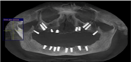
Figure 2: CT scan after 1 month surgery.

Figure 3: Intraoral view of abutments upper jaw before prosthetic reconstruction

Figure 4: Intraoral view of abutments lower jaw before prosthetic reconstruction

Figure 5: Upper and lower jaw implant placement non-removable metalceramic prosthetic restoration before prosthetic reconstruction.

Figure 6: Clinical appearance after prosthetic rehabilitation with non-removable metal-ceramic prosthetic restoration

Figure 7: Clinical appearance after prosthetic rehabilitation with non-removable metal-ceramic prosthetic restoration

Figure 8: CT scan after prosthetic rehabilitation
Discussions
Bone grafting and sinus lifts are invasive procedures. In addition, they add complexity and increase the number of surgical phases required for implant therapy. These techniques pose a series of inconveniences, such as the need for multiple surgical interventions, the use of extraoral bone donor sites (e.g., iliac crest or skull) ‑ with the morbidity involved in surgery of these zones ‑ and the long duration during which patients remain without rehabilitation during the graft consolidation and healing interval [25]. These factors complicate patient acceptance of the restorative treatment and limit the number of procedures carried out.
Sinus lifts procedure is one of the most common preprosthetic surgical procedures performed in dentistry today. However the development and improvement of alternative methods for restoring the integrity of the dentition with an extreme degree of atrophy of the alveolar crest of the upper jaw is very urgent. In clinical practice it is becoming increasingly common for patients to demand therapies that offer a good final result while at the same time reduce costs, healing time and the temporary inability to work. In connection with this, new implant technologies become widespread, which allow to reduce the volume and number of surgical interventions, as well as to shorten the duration of treatment. In order to overcome such limitations, different therapeutic alternatives have been proposed, such as, short implants, or implants placed in specific anatomical areas like the pterygoid region, the tuber or the zygoma [26-30]. Any of these procedures requires considerable surgical expertise and has its own advantages, limits, surgical risks and complications involving biological and financial costs.
The present study shows good clinical outcome with standard implants placed in the tuber regions and zygomatic bone of the maxilla using a two-stage procedure. This offers a more simplified treatment approach, a decrease in biological impact and a more comfortable post-surgical period for the patient thanks to a quicker recovery time. Implants through the atrophied upper jaw in the tuber regions and zygomatic bone of the maxilla are a good alternative to maxillary sinus lift and to bone grafts in patients with posterior atrophic maxillae. These methods allow:
1. Avoid bone grafting, which is used in conditions of atrophy of the upper jaw when installing conventional dental implants.
2. To shorten the terms of rehabilitation: to produce a fully functional and aesthetic prosthesis.
Implantation methods that were used in this work can provide a patient who is not ready for risky, expensive and multi-stage surgical treatment, the possibility of avoiding more traumatic (such as bone transplantation in the patient) and less predictable types of surgical intervention. Carrying out a comparative analysis of the different approaches to the treatment of adentia in patients with severe maxillary atrophy in the area of the maxillary sinus, we came to the conclusion that a reasonable combination of different techniques can be achieved in order to achieve the optimal result. Rehabilitation using implants in the tuber regions and zygomatic bone of the maxilla is a predictable technique, it does not lack in possible complications, and therefore, it should be reserved only to professionals with vast surgical experience, as it requires a long learning curve and prior experience with conventional implants.
Conclusions
With proper case selection, correct indication and knowledge of the surgical technique, the dental implants placement in the tuber regions and zygomatic bone of the maxilla offers advantages in the rehabilitation of severely resorbed maxillae, implants show survival rates (97%) after 5 years. These methods of implantation should be considered as an alternative approach to solving problems arising during prosthetics of the atrophied upper jaw.
Declarations
Conflict of interest and financial disclosure
The author declares that he has no conflict of interest and there was no external source of funding for the present study. None of the authors have any relevant financial relationship(s) with a commercial interest.
Funding
This research did not receive any specific grant from funding agencies in the public, commercial, or not-forprofit sectors.
Ethics approval and consent to participate
This protocol was approved by the Clinical Research Ethics Committee Yerevan State Medical University after M. Heratsi(protocol N3 17.11.16) and in accordance with those of the World Medical Association and the Helsinki Declaration.
Acknowledgements
Not applicable
References
- Alfaro F.N. Bone grafting in oral implantantology techniques and clinical applications Quintessence. 2006; 233 p.
- Khoury F, Antoun H, Missika P. Bone augmentation in oral implantology.Quintessence. 2007;435p.
- Sharan A, Madjar D. Maxillary sinus pneumatization following extractions: A radiografiphic study.IntJ Oral Maxillofac Implants 2008;23(1):48-56.
- Boyne P, Cole M, Stringer D, Shafquat J. A technique for osseous regeneration of deficient edentulous maxillary ridges. J. Oral Maxillofac. Surg 1985; 43:87-91.
- Sailer H.A A new method of inserting endosseous implants in totally atrophic maxillae.JCranio-MaxillofacSurg 1989;17(7):299-305.
- Tonetti MS, Hämmerle CH. European Workshop on Periodontology Group C. Advances in bone augmentation to enable dental implant placement: Consensus Report of the Sixth European Workshop on Periodontology.J ClinPeriodontol 2008; 35: 168-72.
- Rosen A, Gynther G. Implant treatment without bone grafting in edentulous severely resorbed maxillas: A long-term follow-up study. J Oral MaxillofacSurg 2007; 65: 1010-1016.
- Maló P, NobreMde A, Lopes I. A new approach to rehabilitate the severely atrophic maxilla using extramaxillary anchored implants in immediate function: a pilot study. J Prosthet Dent. 2008; 100(5): 354-366.
- Chanavaz M. Sinus graft procedures and implant dentistry: a revien of 21 years of surgical experience (1979-2000). Implant Dentistry.2000; 3: 197-203.
- Tatum, H. Maxillary and sinus implant reconstruction Dent. Clin. North America. 1986; 30: 207-229.
- Misch C.E. The maxillary sinus lift and sinus graft surgery.In: Contemporary implant Dentistry. St Louis: Mosby 1999; 469-495.
- Peleg M, Mazor Z, Chaushu G, Garg AK. Sinus floor augmentation with simultaneous implant placement in the severely atrophic maxilla.JPeriodontol 1988:69:1397-1403.
- Boyne PJ, James RA. Grafting of the maxillary sinus floor with autogenous marrow and bone. J Oral Surg 1980; 38: 613-616.
- Block M.S., Kent J.N. Sinus augmentation for dental implants, the use of autogenous bone. //J Oral MaxillofacSurg 1997; 55:1281-1286.
- Balshi TJ, Wolfinger GJ, Balshi SF. Analysis of 356 pterygo-maxillary implants in edentulous arches for fixed prosthesis anchorage.Int J Oral Maxillofac Implants 1999; 14: 398-406.
- Bidra AS, Huynh-Ba G. Implants in the pterygoid region: a systematic review of the literature.IntJ Oral Maxillofac Surg. 2011; 40(8): 773-781.
- Candel E, Peñarrocha D, Peñarrocha M. Rehabilitation of the atrophic posterior maxilla with pterygoid implants: a review. J Oral Implantol. 2012; 38: 461-466.
- Graves SL. The pterygoid plate implant: a solution for restoring the posterior maxilla.// Int J Periodontics Restorative Dent 1994; 14: 512-523.
- Ridell A., Grondahl K., Sennerby L. Placement of branemark implants in the maxillary tuber region: Anatomical considerations, surgical technique and longterm results. Clinical Oral Implants Research 2009 ;20(1):94-98.
- Jaime G Rodríguez-Chessa, Sergio Olate, Henrique Duque Netto, Jamil Shibli, Márcio de Moraes, and Renato Mazzonetto. Treatment of atrophic maxilla with zygomatic implants in 29 consecutives patientsInt J ClinExp Med. 2014; 7(2):426-430.
- Miglioranca R.M., Coppede, A., Dias Rezende R.C. and de Mayo T. Restoration of the edentulous maxilla using extrasinus zygomatic implants combined with anterior conventional implants: A retrospective study.The International Journal of Oral & Maxillofacial Implants, 2011; 26:665-672.
- Candel-Marti E., Carrillo-Garcia, C., Penarrocha-Oltra, D. and Penarrocha-Diago, M. Rehabilitation of atrophic posterior maxilla with zygomatic implants: Review. Journal of Oral Implantology 2012; 38:653-657
- Malevez C., AbarcaM.mDurdu, F., Daelemans P. Clinical outcome of 103 consecutive zygomatic implants:A 6-48 months follow-up study.Clin Oral Implants Res.2004;115:18-22
- Komagamine Y, Kanazawa M, Minakuchi S, Uchida T, Sasaki Y. Association between masticatory performance using a colour-changeable chewing gum and jaw movement. J Oral Rehabil 2011:38:555-63.
- Iturriaga MT, Ruiz CC. Maxillary sinus reconstruction with calvarium bone grafts and endosseous implants. J Oral Maxillofac Surg. 2004; 62(3):344-347.
- José-Luis Sierra-Sánchez et al. Predictability of short implants (< 10 mm) as a treatment option for the rehabilitation of atrophic maxillae. A systematic review.Med Oral Patol Oral Cir Bucal.2016 ; 21(3): 392-402.
- Reiser GM. Implant use in the tuberosity, pterygoid, and palatine region anatomic and surgical considerations. In: Nevins M, Mellonig JT (eds) Implant therapy clinical approaches and evidence of success, 2nd edn. Quintessence Books, Chicago,1998.
- N., Girish Rao. Pterygomaxillary Implants: A Graftless Solution to Deficient Maxillary BoneJ Indian Prosthodont Soc. 2012; 12(3): 182-186.
- Prithviraj R.V., Harleen K.B. From maxilla to zygoma: A review on zygomatic implants.Journal of Dental Implants 2014;4(1):44-47
- Galán-Gil S, Penarrocha-Diago M, Balaguer-Martνnez J, Marti-Bowen E. Rehabilitation of severely resorbed maxillae with zygomatic implants: an update.Med Oral Patol Oral Cir Bucal. 2007;12(3):216-220